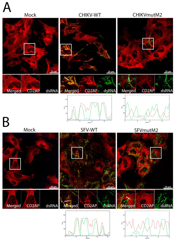
Figure 7.
dsRNA in wt CHIKV and SFV infected cells co-localisation with CD2AP. HOS cells infected with CHIKV wt or CHIKVmutM2 (A) or SFV wt or SFVmutM2 (B) at an MOI of 1 were fixed at 16 (A) or 8 h pi (B). Thereafter the cells stained for dsRNA (green) and CD2AP (red), line scan image is given below. Results are representative of three independent experiments.

