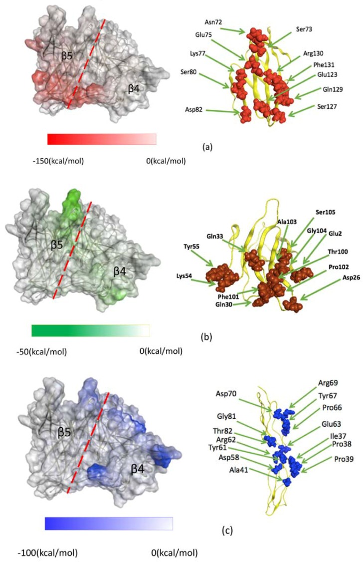
Figure 3.
Interfaces of MVH and the respective three receptors. (Left) β4 and β5 strand regions on the MVH surface that show strongly attractive interactions with the three receptors: (a) SLAM (red), (b) Nectin-4 (green), and (c) CD46 (blue). The deepness of the color represents the strength of the IFIE sum associated with the fragment (MVH residue) at that position. The dashed line represents the approximate boundary between the β4 and β5 regions. (Right) Residues on the receptor surface that show importantly attractive interactions with MVH: (a) SLAM, (b) Nectin-4, and (c) CD46.

