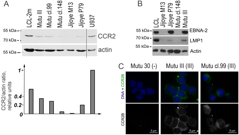
Figure 3.
CCR2 protein expression in the EBV-positive Burkitt lymphoma (BL) cell lines. Immunoblotting analyses of BL cell lines Mutu III (latency III), Mutu cl.99 (latency III), Mutu cl.148 (latency I), Jijoye M13 (latency I), Jijoye P79 (latency III) for detection of the CCR2 protein (A) and the EBV antigens EBNA2 and LMP1 (B). The LCL cell line with latency III (LCL-2m) and the human myeloid histiocytic lymphoma cell line U937 are used as the positive controls. (C) CCR2B was determined by immunofluorescence microscopy using immunostaining with the mouse monoclonal anti-CCR2B antibody. The nuclear DNA staining with Hoechst is shown in blue. Mutu 30 (-) is the EBV-negative BL line Mutu 30. Scale bar = 5 μm.

