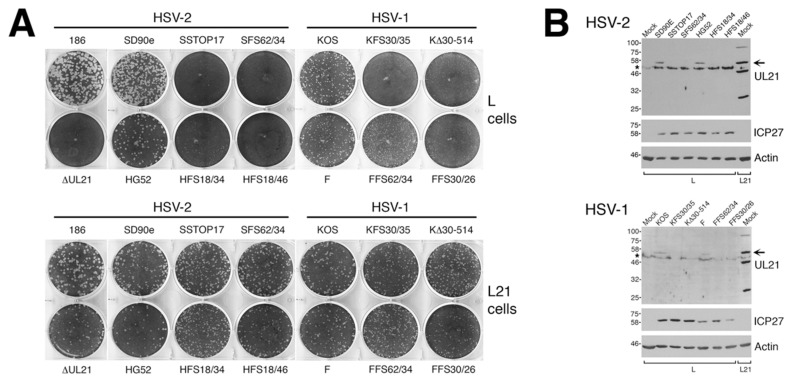
Figure 2.
Confirmation of pUL21 deficiency in CRISPR/Cas9 mutants. (A) Monolayers of L cells or L21 cells were infected with equivalent numbers plaque forming units of the indicated viruses, overlaid with semisolid medium containing 1% methyl cellulose, then fixed and stained with 70% methanol containing 0.5% methylene blue at 72 hpi. (B) Western blot analysis of CRISPR/Cas9 mutants. Whole cell lysates prepared at 24 hpi from L cells infected at a multiplicity of infection (MOI) of 0.1 with the indicated viruses, or from mock infected L or L21 cells, were separated on SDS 8% polyacrylamide gels and transferred to membranes. Membranes were blocked overnight at 4 °C, then probed for the presence of pUL21, ICP27 (infection control), or actin (loading control). Positions of protein molecular weight markers in kDa are indicated on the left. Horizontal arrows indicate the position of full-length pUL21. Asterisks on the left side of the pUL21 panels denote the position of a cellular protein that cross reacts with the pUL21 antiserum.

