Figure 3.
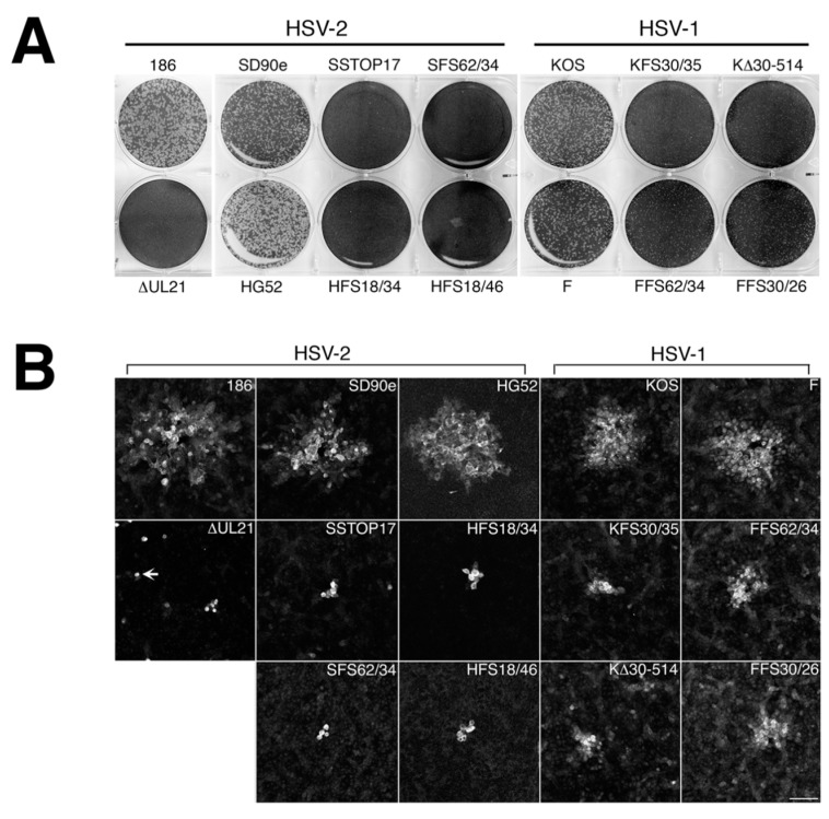
Cell-to-cell spread capabilities of HSV UL21 deletion mutants. (A) Monolayers of Vero cells were infected with equivalent numbers plaque forming units of the indicated virus strains, overlaid with medium containing 1% methyl cellulose, then fixed and stained with 70% methanol containing 0.5% methylene blue at 72 hpi. (B) Monolayers of Vero cells growing on glass bottom dishes were infected with the indicated viruses and overlaid with medium containing 1% methyl cellulose. At 24 hpi, infected monolayers were fixed, permeabilized, and then stained for the presence of the HSV kinase Us3. Representative images are shown. Arrow indicates a single infected cell, which were very prominent in Vero cells infected with ∆UL21 at 24 hpi. Scale bar is 100 μm. (C) The plaque areas of 40 plaques per virus strain from the experiment depicted in (B) were measured using Image-Pro 6.3 software (Media Cybernetics, Bethesda, MD, USA). Single infected cells were not included in this quantification. Error bars are standard error of the means.


