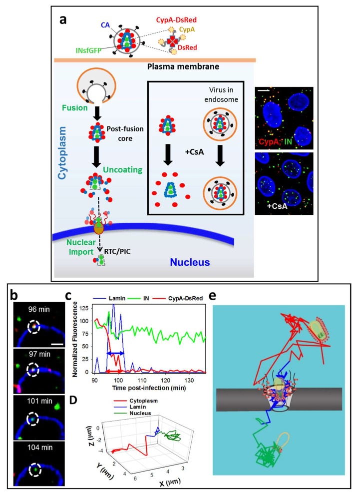
Figure 2.
A novel capsid protein (CA) marker CypA-DsRed enables visualization of single HIV-1 uncoating. (a) Illustration of HIV-1 co-labeling with INsfGFP that marks the viral pre-integration complexes and the tetrameric CA marker, CypA-DsRed (inset), that remains bound to CA after viral fusion. Single HIV-1 uncoating and nuclear import are shown. The second advantage of this labeling strategy (shown boxed) is the ability to identify post-fusion cores by Cyclosporine A (CsA) treatment. CsA selectively displaces CypA-DsRed from post-fusion cores, but not from intact viruses trapped in endosomes; (b–e) Uncoating and nuclear import of single HIV-1 cores in TZM-bl cells. Confocal images (b) and fluorescence intensity traces (c) of uncoating and nuclear import are shown; (d) Single particle trajectory corrected for the nucleus movement. Segments of the trajectory are colored to mark the cytoplasmic transport, docking and intra-nuclear movement. Dotted circles in (b) mark a single viral complex uncoating and entering the nucleus. Double arrows in (c) illustrate the virus docking time (colocalization with the lamin signal, blue) and the time of uncoating after docking (red). A model for HIV-1 uncoating and nuclear import is overlaid onto an actual 2D-trajectory of single HIV-1 undergoing uncoating and nuclear import (e). Scale bar 5 µm in (a) and 2 µm in (b). Adapted from Francis et al., 2016 and Francis and Melikyan, 2018.

