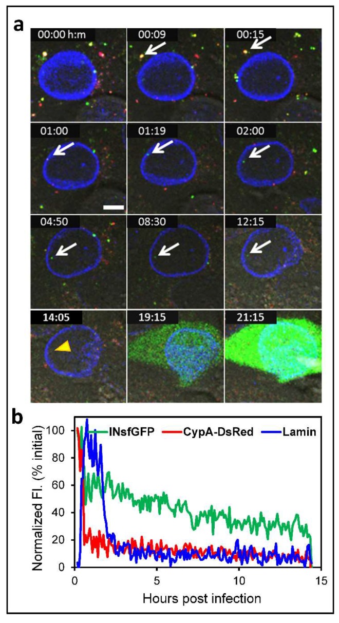
Figure 3.
Live cell imaging of single HIV-1 uncoating and nuclear import that culminate in infection. TZM-bl cells expressing EBFP2-Lamin were infected with VSV-G pseudotyped HIV-1 encoding for the eGFP reporter. Viruses were labeled with the INsfGFP and CypA-DsRed markers. Confocal time-lapse images were acquired every 5 min from 0 to 24 h post-infection. (a) Time-lapse images of single HIV-1 entry and infection. The arrow marks a single INsfGFP labeled HIV-1 complex in the cytoplasm that docks at the NE, uncoats, and enters the nucleus and disappeared at 14:05 hours post-infection (yellow arrowhead). Loss of INsfGFP signal is followed by expression of eGFP reporter of infection. Scale bar 5 µm. (b) Fluorescence intensity traces of the virus in (a) that undergoes terminal uncoating after engaging the EBFP2-Lamin labeled NE (manifested in the increase in lamin signal) and enters the nucleus (drop in the lamin signal). Single particle tracking was performed using INsfGFP as reference. The drop in the INsfGFP signal at ~14 h post-infection marks disappearance of the IN spot prior to eGFP expression. Scale bar 5µm.

