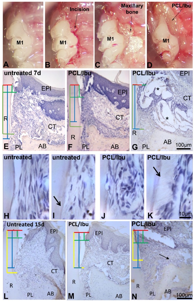
Figure 2.
Periodontitis induced with Porphyromonas gingivalis-infected ligatures and treatment with PCL/Ibu membrane (A–D). (B) sulcular incision along the first and second maxillary molars, (C) raising the flaps for exposure and access, (D) surgical placement of PCL/Ibu membrane on the periodontal lesion, (E–N) histological view at 7 and 15 days. (E–K) histology of periodontal wound healing at 7 days and (L–N) at 15 days. Red line = cementoenamel junction, blue line = fibrous connective tissue attachment, green line = epithelial attachment, yellow line = bone level. After anesthesia, a slight incision to the bone crest contact was made to facilitate the first ligature placement at the junction between the gum and the tooth along the first and second molars (M1-M2) as previously described [16]. The thread was then blocked with a drop of glass ionomer (Fuji IIGC, GC, France, Bonneuil sur Marne, France). Sterilized black braided 6.0 silk threads (Ethicon, Auneau, France) were incubated in culture medium containing P.gingivalis in an anaerobic chamber for one day. P.gingivalis-soaked ligatures were placed around maxillary first and second molars. The ligatures were inspected and replaced (with freshly infected ones) thrice a week for a period of 40 days. An incision was performed along the sulcular margins of the first and second molars and extended anteriorly on the mesial aspect of the first molar to efficiently raise the flap to gain access. Ibuprofen-functionalized PCL membrane was punched with a 3 mm diameter cutter. The circular pieces of membrane were further divided into half to achieve a size appropriate enough to cover the lesion. The cut membrane was then placed into the periodontal pocket after raising the flap such that the membrane stays flat beneath the flap covering the lesion fully and the necks of the crowns (molars) partially, entering the inter-dental area as well. The flap was nicely repositioned to perform a suture on the flap while maintaining the membrane underneath [16]. AB: alveolar bone, CT: connective tissue, EPI: epithelium, PL: periodontal ligament, R: root. Stars showing PCL/Ibu membrane.

