Figure 3.
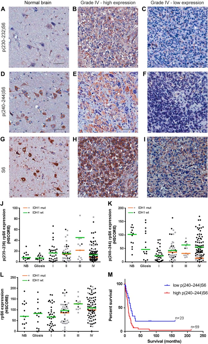
rpS6 expression and phosphorylation in gliomas according to grade and IDH1 status. Tissues of non-tumoral brain (NB), reactive gliosis (gliosis), and astrocytomas of grades I–IV (pilocytic: I, diffuse: II, anaplastic: III, and GBM: IV) were spotted into a TMA, and immunohistochemistry reactions were performed for p(232–235)S6, p(240–244)S6, total S6, and IDH1 R132H. Representative immunohistochemistry of non-tumoral brain (A, D, and G) as well as GBMs with high (B, E, and H) and low (C, F, and I) S6 phosphorylation at 232–235 and 240–244, respectively. Reactions were quantified automatically, and the HSCORE was calculated considering the intensity of labeling and the number of labeled cells for the phosphorylation of residues 232–235 (J), 240–244 (K), and total expression (L). Samples positive for IDH1 R132H were classified as mutant (mut), and samples negative for IDH1 R132H were classified as wild-type. The overall survival curve of GBM patients according to the levels of S6 phosphorylation at residues 240–244 is also shown (M). The cutoff for classification of the sample between high and low was the first quartile (HSCORE 45). Not statistically different from NB, p<0.05. Scale bar 50 µm. Abbreviations: NB, non-tumor brain; GBM, glioblastoma; TMA = tissue microarray.

