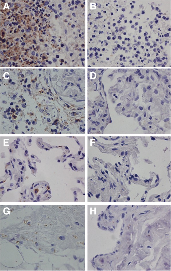
Fig. 3.

Immunohistochemistry results of a deceased SFTSV patient. The spleen tissues had the most amounts of SFTSV antigens (Panel a); the kidney had moderate amount of SFTSV antigens (Panel c), and the lung (Panel e) and the heart (Panel g) had least amount SFTSV antigens. Negative controls were stained by omitting the primary antibody incubation using the spleen (Panel b), kidneys (Panel d), lungs (Panel f), heart (Panel h) tissue sections respectively. (original magnification × 400)
