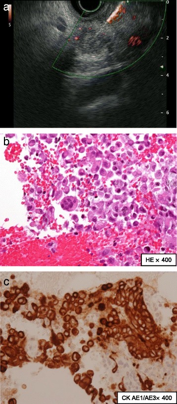
Fig. 2.

Findings obtained by endoscopic ultrasound-guided fine-needle aspiration. a Endoscopic ultrasound-guided fine-needle aspiration was performed to obtain cytology for the solid mass in the pancreatic head. b Histology showed pleomorphic large atypical cells (hematoxylin-eosin, magnitude × 400). c Cytokeratin AE1/AE3 stain was positive, and thus these cells were epithelial cells (cytokeratin AE1/AE3, magnitude × 400)
