
Figure 6.
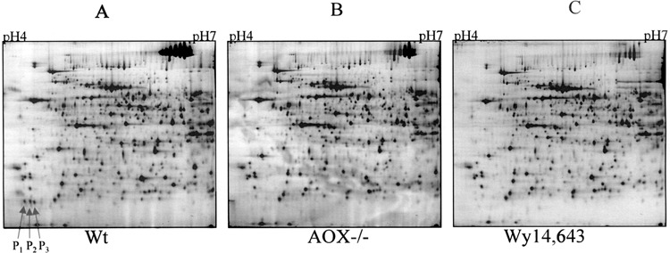
2D gel electrophoresis of mouse liver proteins. Mouse liver homogenates of wild-type (A), AOX−/− (B), and wild-type treated with Wy-14,643 (C) were first separated by isoelectric focusing (horizontal axis, pH 4–7) and further separated by 10% SDS-PAGE (vertical axis), which stretches from approximately 15 kDa (bottom) to about 120 kDa (top). Arrows indicate the PP-downregulated 18.7-kDa triple spots.