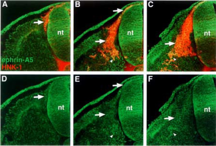
Figure 3.

Ephrin-A5 is most strongly expressed by neural crest cells that have delaminated and migrated ventrally in the sclerotome. (A, B, C) Cross sections through stages 14–18 embryos stained with ephrin-A5 antibody (green) and HNK-1 antibody (red), a marker for avian neural crest. (D, E, F) Ephrin-A5 antibody labeling. Ephrin-A5 localizes predominantly to recently delaminated neural crest prior to somite entry (A–F: arrows), to migratory neural crest (B, E: arrows), and to neural crest at ventral locations in the somite (B, C, E, F: arrowheads).
