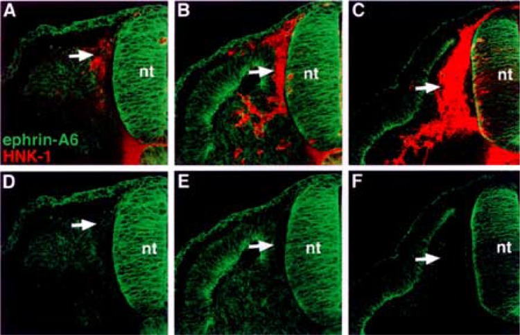
Figure 4.

Ephrin-A6 protein associates with neural crest cells prior to their entry into the somite. (A, B, C) Cross sections through stages 14–18 embryos stained with ephrin-A6 antibody (green) and HNK-1 antibody (red). (D, E, F) Ephrin-A6 antibody labeling. Ephrin-A6 protein is evident on recently delaminated neural crest that lie between the neural tube and somite (A, D: arrows). As neural crest cells enter the somite, ephrin-A6 protein is not detectable (B–F: arrows).
