
Figure 5.
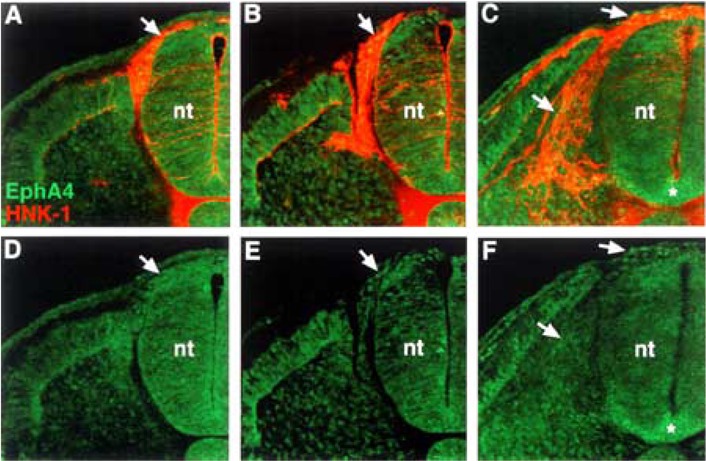
EphA4 protein is present at very low levels on neural crest cells. (A, B, C) Cross sections through stage 18 embryos, at different trunk axial levels, stained with EphA4 antibody (green) and HNK-1 antibody (red). (D, E, F) EphA4 antibody labeling. EphA4 protein is apparent at very low levels on neural crest that have exited the neural tube and are migrating in the somites (arrows). Asterisk (*) marks EphA4 expression in the floor plate.