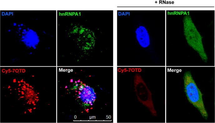
Figure 7.

Fluorescence microscopy images of HeLa cells treated with Cy5–7OTD and then immunostained with antibodies to hnRNPA1. G-quadruplex RNA was detected by Cy5–7OTD (red). hnRNPA1 was immunostained using antihuman hnRNPA1 antibodies (green). Blue fluorescence indicates foci derived from DNA stained by DAPI. The merged panel shows a colocalization event. RNase-treated cells show a greatly reduced RNA signal.
