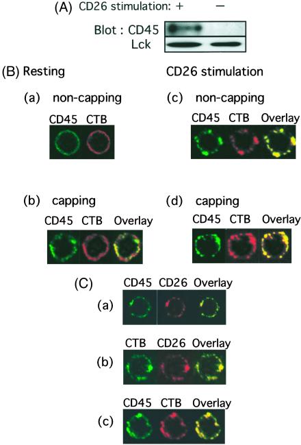
Figure 5.
Colocalization of CD26 and CD45 in lipid rafts after modulation of CD26. (A) Unstimulated or 1F7-stimulated T cells were subjected to sucrose gradient centrifugation to separate lipid raft fractions from Triton-soluble fractions. Protein contents of fraction 4 of the sucrose gradient were resolved by SDS/PAGE, and protein patterns were analyzed on immunoblots stained with the corresponding Abs. CD45 (UCHL1) was enriched in rafts after CD26 stimulation. (B) Following staining with anti-CD45 (FITC-conjugated UCHL1) and CTB (biotinylated), cells were then analyzed for potential colocalization of CD45 and CTB patches. After staining of these molecules on the cell surface with (b and d) or without (a and c) capping, CD26 was crosslinked and internalized with 1F7 mAb (c and d). (C) Cells were stained by the biotinylated anti-CD26 antibody 5F8 followed by Texas red-conjugated streptavidin and the FITC-conjugated anti-CD45RO antibody UCHL1 (a), FITC-conjugated CTB and biotinylated 5F8 (b), or FITC-conjugated UCHL1 and biotinylated CTB (c). After staining of these molecules on the cell surface, CD26 was crosslinked and internalized with 1F7 mAb.

