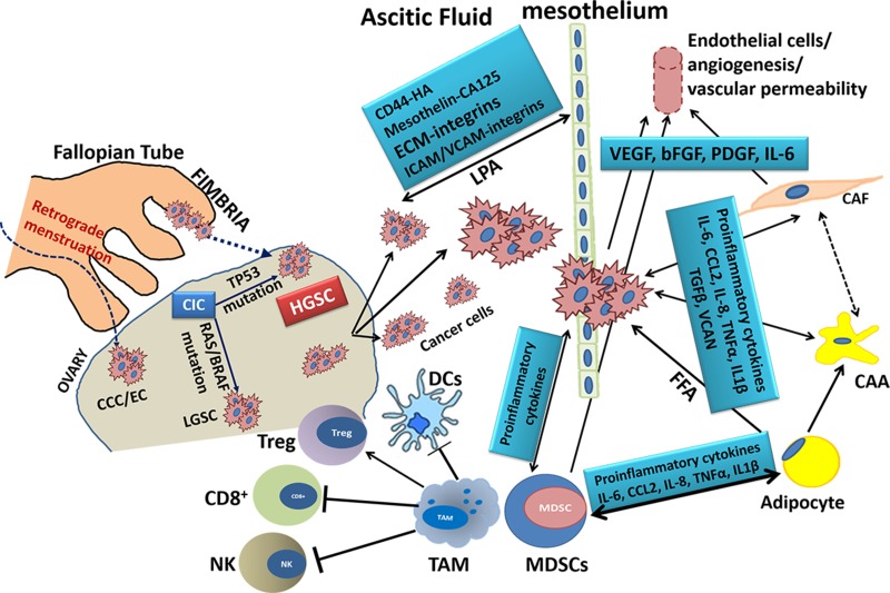
Figure 1. Schematic Representation of the key cell types in Ovarian Cancer Microenvironment and the molecules involved in their interactions.
HGSC: high grade serous cancer; LGSC: low grade serous cancer; CCC: clear cell carcinoma; EC: endometrial carcinoma; CIC: carcinoma in situ; CAA: cancer-associated adipocyte; CAF: cancer-associated fibroblast; FFA: free fatty acids; VEGF: vascular endothelial growth factor; bFGF: basic fibroblast growth factor; PDGF: platelet-derived growth factor; VCAN: versican; CD8+, cytotoxic T cell; Treg: regulatory T cell; DCs: dendritic cells; MDSCs: myeloid-derived suppressor cell; ECM, extracellular matrix; IL-x, interleukin-x; ICAM/VCAM: intercellular/vascular adhesion molecule; HA: hyaluronic acid; CA125: cancer antigen 125; LPA: lysophosphatidic acid; NK: natural killer cell; TAM: tumor-associated macrophage; TGFβ: growth transforming growth factor β; TNFα: tumor necrosis factor-α.

