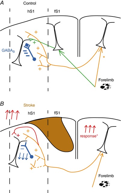
Figure 8. Proposed model of the synaptic changes occurring in callosal connectivity in the periphery of a focal stroke.

A, normal condition: callosal inputs (orange pathway) inhibits sensory responses mediated by the direct activation of the contralateral limb (green pathway) via activation of locally residing interneurons that act via GABAB (blue). *Under normal conditions limb tactile stimulation activates the ipsilateral cortex (e.g. (Takatsuru et al. 2009); Kokinovic and Medini, unpublished observation). B, after a focal stroke, recovery of forelimb responses cannot occur via horizontal connections from the stroked area. Instead, recovery happens via the combined effect of reduced strength of IHI (blue) and by potentiation of the direct callosal excitatory input (red). The latter can be accounted for increased strength of the local excitatory synapses present in the stroke periphery and/or by an increased activation of the contralesional cortex (Reinecke et al. 2003; Takatsuru et al. 2009). [Color figure can be viewed at http://wileyonlinelibrary.com]
