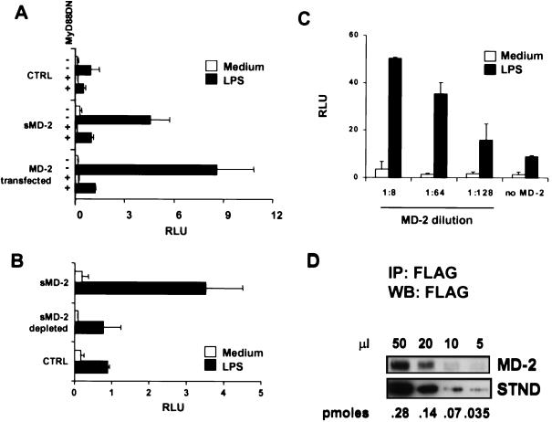
Figure 2.
sMD-2 confers LPS responsiveness to TLR4+/MD-2− reporter cells. (A) 293TLR4 cells were cotransfected with NF-κB and β-Gal reporter vectors and either dominant negative MyD88 (MyD88DN) (+) or empty vector (−). After 24 h, each group of transfectants was treated with culture medium from 293 (CTRL) or 293MD-2 cells (sMD-2) in the presence or absence of LPS. As a comparison, 293TLR4 cells that had been transfected with MD-2 were treated with or without LPS (MD-2 transfected) in fresh medium. Bars indicate relative NF-κB activity. Data are representative of six separate experiments. (B) The LPS-enhancing activity is depleted by anti-FLAG mAb. 293TLR4 cells were left untreated (open bars) or treated with LPS (filled bars) in medium alone (CTRL), in medium containing supernatant from 293MD-2 cells (sMD-2), or in 293MD-2 cell medium adsorbed with anti-FLAG mAb (sMD-2 depleted). (C) Dose-response of MD-2. 293TLR4 cells were incubated with the indicated dilutions of concentrated 293MD-2 culture medium or with medium alone (no MD-2), and the LPS-dependent activation of NF-κB was followed as in A. (D) Quantitation of sMD-2. Serial dilutions of concentrated 293MD-2 culture medium (MD-2) were immunoprecipitated with the anti-FLAG mAb, resolved by SDS/10% PAGE, blotted with HRP-anti-FLAG, and developed by enhanced chemiluminescence. Intensities were compared with those from known concentrations of a standard protein (STND, carboxyl-terminal FLAG-BAP fusion protein, Sigma) shown in the lower panel. By comparing intensities, we estimate that 20 μl of the MD-2 concentrate contained ≈0.1 pmol of MD-2.

