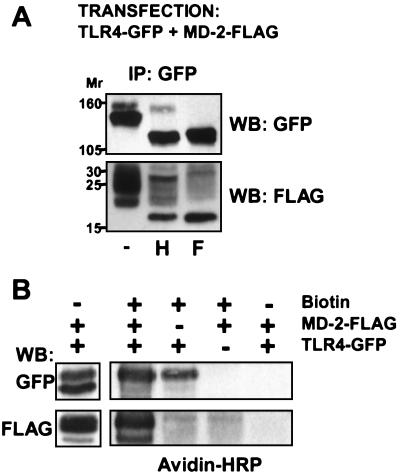
Figure 6.
TLR4 and MD-2 associate in the cytoplasm. (A) 293T cells were cotransfected with TLR4-GFP and MD-2-FLAG, and lysates were immunoprecipitated after 48 h with the anti-GFP pAb and protein A Sepharose. Beads were left untreated (−) or treated with endo H (H) or PNGase F (F), solubilized in SDS reducing buffer, and analyzed by Western blotting after 6% (upper membrane) or 13% (lower membrane) SDS/PAGE. The upper membrane was blotted for TLR4, the lower membrane for MD-2. This experiment has been repeated twice with similar results. (B) 293T cells were transfected with either TLR4-GFP or MD-2-FLAG or both, as indicated, and cell-surface proteins were modified (or not) with sulfosuccinimidobiotin. Cells were lysed, immunoprecipitated with the anti-GFP, and analyzed by SDS/PAGE and Western blotting using either anti-GFP or anti-FLAG antibodies (left lane) or HRP-avidin (right lanes).

