
Figure 9. HUVEC Adhesion Site Formation on Multifaceted Patterned Surfaces.
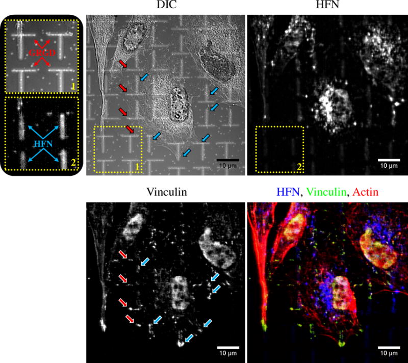
Multifaceted patterned surfaces presenting 1×8 μm ellipses of GRGD-terminated alkanethiol in the horizontal direction (1% GRGD in OEG: see inset 1) interwoven with HFN ellipses in the vertical direction (25 μg ml−1: see insets 1 and 2) were prepared with LSL. The GRGD and HFN patterns displayed an RGD surface density of ~45,165 and 4,593 RGD μm−2 respectively (see Fig.8). To determine the placement of the HUVEC adhesion sites and which ligand was being ligated, the HUVECs were fixed 16 hr post seeding, immunolabeled for HFN (top right), vinculin (bottom left), and actin (red in bottom right), and imaged with fluorescent and differential interference contrast (DIC) (top left) microscopies. HUVECs displayed adhesions to both the GRGD and HFN patterns. The red arrows in the DIC and vinculin images depict cell adhesions formed on GRGD patterns while the blue arrows indicate those formed on HFN patterns. The data demonstrates that both ligands were biologically active after patterning and induced integrin ligation and adhesion site formation. SB=10 μm.