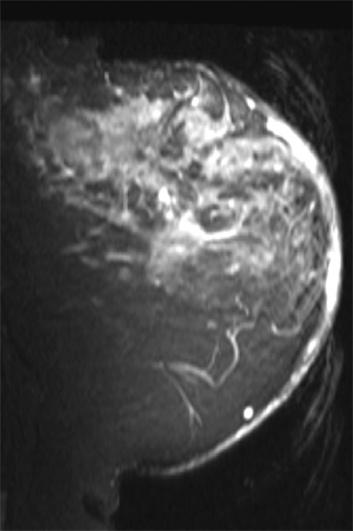
Figure 3a:

Images in a 61-year-old postmenopausal woman with a moderately differentiated invasive lobular carcinoma show a patient with no response (RCB-III) to neoadjuvant chemotherapy (NAC). With MR imaging, it was determined that she had a large tumor of 9.5 cm in diameter at the start of the therapy. She did not respond well to NAC and was classified as RCB-III at the time of surgery. Image a displays an axial MR image obtained prior to chemotherapy. Image b are sagittal diffuse optical tomography images obtained from the left and right breasts just before therapy. The images displayed refer to a time point of 15 seconds after the breath hold and show the percentage change in deoxyhemoglobin (%Δ[Hb]). Finally, image c shows time-dependent signal traces in response to a 30-second breath hold for the tumor region obtained from dynamic diffuse optical tomography just before the start of NAC (baseline) and 2 weeks after treatment initiation.
