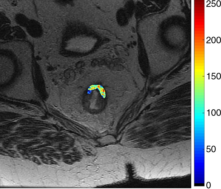
Figure 4b:

Images show radiomics analysis in a 60-year-old man with rectal adenocarcinoma after completion of chemotherapy–radiation therapy with surgically proven complete response. (a) High-spatial-resolution axial oblique T2-weighted image demonstrates treated area with low signal intensity (arrow). (b–e) Illustration of intensity-based texture features overlaid on high-spatial-resolution axial T2-weighted images as follows: (b) contrast, (c) Gabor 45 contrast, and (d) homogeneity.
