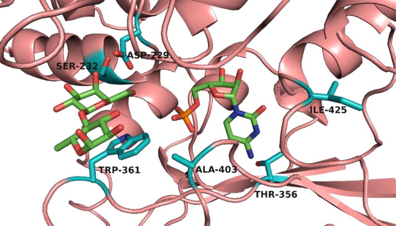
Figure 1.
The substrate binding site of Δ15Pd2,6ST(N) structure modelled based on the co-crystal structure of Δ16Psp2,6ST (PDB ID: 2Z4T) with CMP and lactose (represented with green-colored carbons). Structural modelling was performed with SWISS-MODEL. The six sites chosen for mutagenesis are represented with teal-colored carbons.

