
Fig. 2.
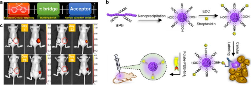
(a) Donor-bridge-acceptor structure of SPs. (b) Schematic diagram showing the preparation of carboxylic-acid-functionalized SPN9 and conjugation with biomolecules for the specific cellular targeting and in vivo bioimaging. (c) Time-dependent in vivo bright-field (left) and fluorescence (right) imaging of a mouse implanted SKOV-3 cells labeled with bare SPN9 (left black circle) or folic acid functionalized SPN9 (right red circle). Reprinted with permission from Ref. [80], copyright 2017, The Royal Society of Chemistry.