
Fig. 4.
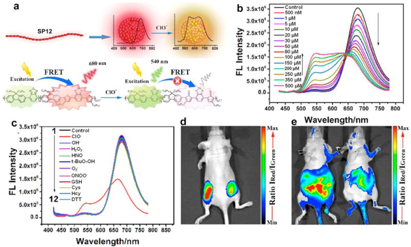
(a) Schematic illustration of SPN12 preparation by nanoprecipitation and ClO− sensing. (b) Fluorescence performance of SPN12 in the presence of different concentrations of ClO−. (c) Fluorescence spectra of SPN12 in the presence of various ROS and biologically relevant analytes. (d) In vivo imaging of exogenous ClO− using SPN12. Representative ratiometric (pseudocolor) image of mouse with the subcutaneous implantation of SPN12 (left) and SPN12 + ClO− (right). (e) In vivo imaging of endogenous ClO− production from the peritoneal cavity of the mice with SPN12 during an LPS-mediated inflammatory response. Representative ratiometric images (pseudocolor) of mice intraperitoneally treated with saline (left) and LPS (right), followed by an intraperitoneal injection of SPN12 at 4 h later. Fluorescence images were acquired 30 min after the injection of SPN12 with a 465 nm excitation filter and 540 nm (green channel) and 680 nm (red channel) emission filters. Ratiometric images were obtained by pixel-by-pixel calculation using ImageJ software. Reprinted with permission from Ref. [87], copyright 2017, American Chemical Society.