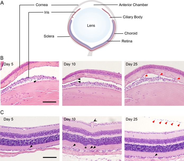
Fig 2. MCMV infection causes inflammation in the iris and retina.
(A) Schematic diagram with the major compartments of the eye labelled. (B) Sections of the anterior chamber at the indicated times pi were stained with haematoxylin and eosin. Thickening of the iris was evident at day 5. Cellular infiltrates and keratic precipitates were present after infection (black arrows). Synechia (adherence of the iris to the cornea) was frequently observed (red arrows). (C) Haematoxylin and eosin stained sections of the retina show normal structure at day 5 pi. Enlarged vessels (two tailed arrow head), folds in the retina (asterisk) and infiltrating cells were present (black arrows) at day 10pi. At day 25 pi infiltrating cells within the retina (black arrow heads) and the vitreous (red arrow heads) were evident. Scale bar: 100 μm. Results are representative of those from 5 mice per time-point.

