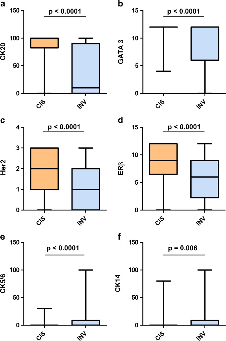
Fig. 2.
Marker expression of CIS and concomitant invasive tumor. a Cytokeratin (CK) 20 (percentage of positive cells evaluated). b GATA3 (Remmele Score). c Human epidermal growth factor receptor 2 (Her2) (DAKO Score). d Estrogen Receptor (ER) β (Remmele Score). e CK5/6 (percentage of positive cells). f CK14 (percentage of positive cells). Band indicates median, bottom, and top of box show first and third quartiles, whiskers demonstrate range of data distribution (minimum/maximum). A significant downregulation of luminal markers and an upregulation of basal markers were observed in the invasive compartment

