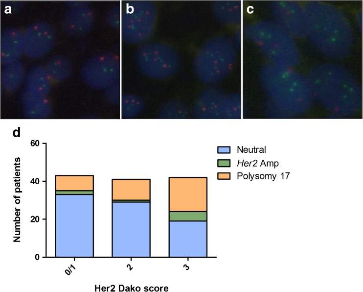
Fig. 3.
Fluorescence in situ hybridization (FISH). a Neutral (non-amplified, non-polysomic). b Polysomy 17. c Her2 amplification. d Distribution of Her2-amplified (Amp), chromosome 17 polysomic, and neutral cases among the various Her2 protein-staining intensities, measured by Dako score. Red hybridization signals indicate the centromeric region of chromosome 17; green signals bind to the Her2 gene locus on chromosome 17

