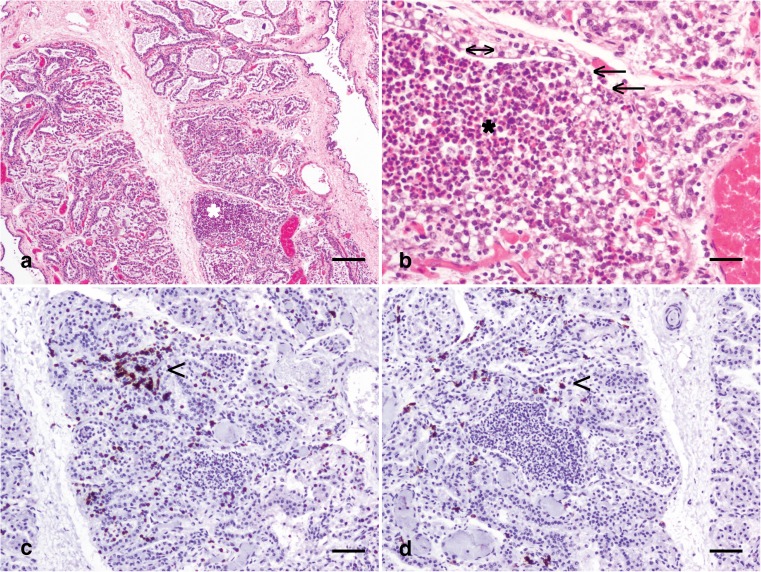
Fig. 1.
The mammary microenvironment in mastitis in a third lactation Holstein Friesian cow, 46 dL. (a) Multifocal mammary alveoli are engorged with numerous predominantly degenerate neutrophils (*). Haematoxylin and eosin stain; scale bar: 300 μm. (b) Severely affected alveoli with myriad neutrophils (*) exhibit partial loss of the luminal epithelial lining (arrows) although the partial remnants of the mammary epithelial lining remain (double headed arrow). Haematoxylin and eosin stain; scale bar: 50 μm. (c) Scattered aggregates of lymphocytes expressing CD3 (arrowhead) are present multifocally. Immunohistochemical staining for CD3 with haematoxylin counterstain; scale bar: 100 μm. (d) Rarer individual lymphocytes expressing CD20 (arrowhead) are present between mammary alveoli. Immunohistochemical staining for CD20 with haematoxylin counterstain; scale bar: 100 μm. dL: days lactation

