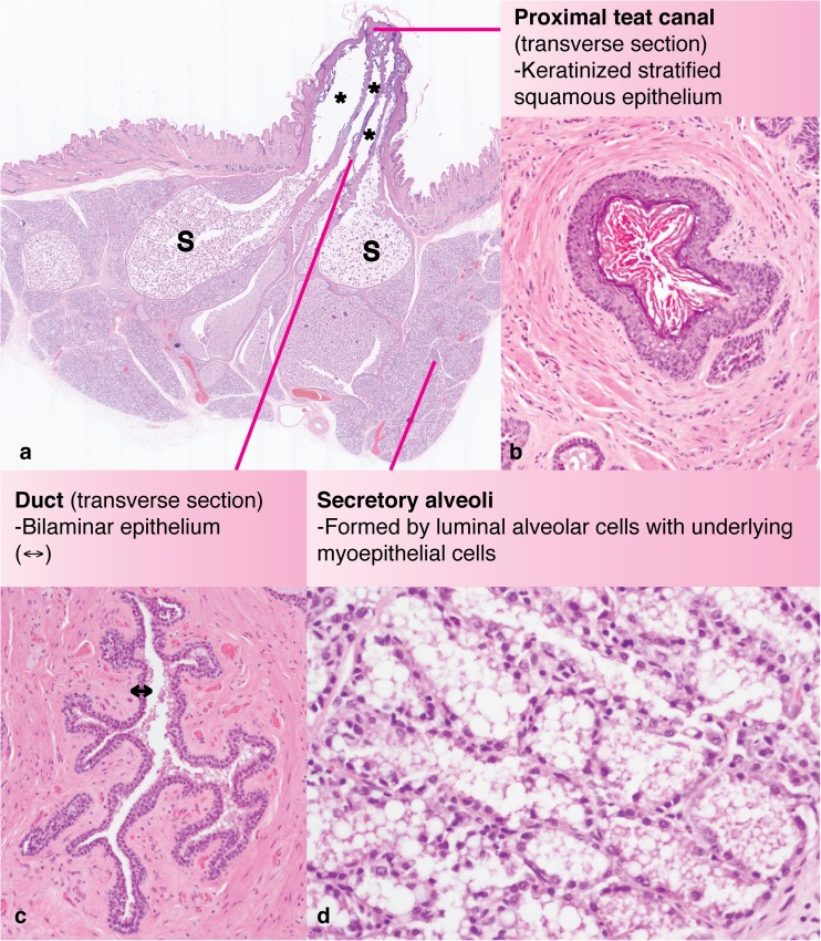
Fig. 2.
Sub-gross anatomy and histology of the rabbit mammary gland. (a) Sub-gross histological section (sagittal plane) through the teat and mammary tissue of a wild rabbit, Oryctolagus cuniculus, during late pregnancy, estimated 27 dG. Multiple ducts (*) are apparent and exhibit dilatations suggestive of sinusoidal structures (S). (b) Transverse section of a rabbit teat canal, < 1 mm from the teat orifice, demonstrating the keratinized stratified squamous epithelium. (c) Transverse section of a mammary duct demonstrating the bilaminar epithelial lining (double headed arrow). (d) Mammary alveoli formed by a luminal layer of mammary epithelial cells and an underlying layer of myoepithelium. Haematoxylin and eosin stain; dG: days gestation

