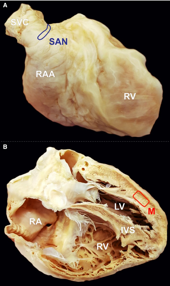
Figure 1.

Photographic image of cadaveric heart specimen with two marked regions from where the samples were obtained: (A) sinoatrial node (SAN) area, (B) left ventricle free wall. IVS, interventricular septum; LV, left ventricle; M, working cardiomyocyte sample; RA, right atrium;RAA, right atrial appendage; RV, right ventricle; SVC, superior vena cava.
