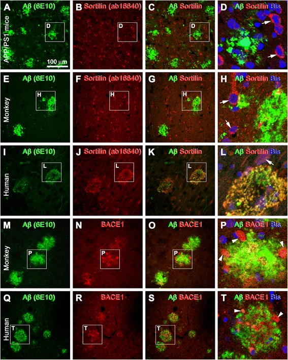
Fig. 5.

Double-immunofluorescence characterization of sortilin labeling relative to amyloid plaque pathology in transgenic mouse, aged monkey, and human neocortex. Species, antibodies, and fluorescence channels are indicated. The framed areas are enlarged as the last panel for each set of double-immunofluorescence images, with bisbenzimide (Bis) nuclear stain included (d, h, l, p, t). a–d Double-immunofluorescence with the rabbit anti-sortilin and 6E10 antibodies in the parietal neocortex of the amyloid precursor protein and presenilin 1 double-transgenic (APP/PS1) model (14 months of age) as an example of the lack of extracellular sortilin labeling in transgenic mouse forebrain with β-amyloid (Aβ) deposition. e–h The same situation as above in aged monkey neocortex (from a 34-year-old rhesus monkey). Note that neuronal somata and proximal dendrites (white arrows) show distinct sortilin immunolabeling in both the transgenic (b, d) and monkey (f, h) cortex. i–l Colocalization of sortilin and Aβ immunofluorescence in two circular plaques in human cortex (positive control). m–t Neuritic plaques in the monkey (m–p) and human (q–t) cortex consisted of extracellular Aβ deposits intermingling with dystrophic neurites (P and T, white arrowheads) exhibiting strong β-secretase 1 (BACE1) immunoreactivity. Scale bar = 100 μm in (a) applying to (b, c, e-g, i–k, m–o, q–s), equivalent to 33 μm for (d, h, l, p, t)
