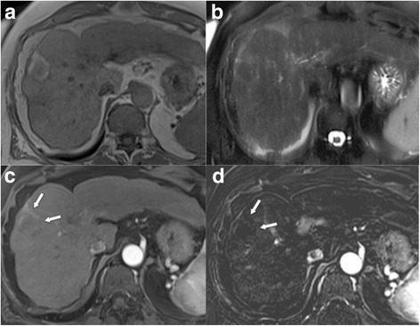
Fig. 14.

LR-TR nonviable a T1w GRE image after tumour ablation demonstrates hyperintense necrosis zone, which is b hypointense on T2w TSE image. c Contrast-enhanced arterial phase images shows absence of lesion enhancement (arrows). d However, this best seen on the subtracted arterial phase image (arrows)
