
Fig. 9.
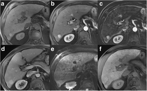
LR4. a Unenhanced T1w GRE image reveals a 17 mm nodule in the right lobe, which is b hypervascular in the arterial phase, best seen c in the subtraction image. d There is no washout present in the venous phase. e Ancillary features favouring malignancy are restricted diffusion in DWI and f hypointensity in hepatobiliary phase. According to size and hypervascularity without washout, lesion would be categorized as LR-3, but ancillary features justify upgrade to LR-4. Biopsy revealed HCC