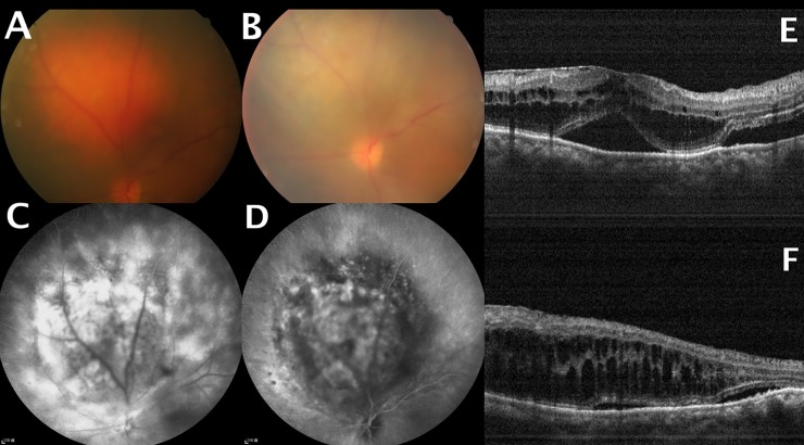
Fig 1.
(A) Pretreatment fundus photograph showing a choroidal hemangioma touching the optic disc. (B) Fundus photograph revealed partial tumor regression after five photodynamic therapy (PDT) sessions. (C) Pretreatment fluorescein angiography demonstrated tumor hyperfluorescence. (D) Washout effect on indocyanine angiography. (E) Pretreatment optical coherence tomography (OCT) showing subfoveal fluid accumulation and cystoid macular edema. (F) OCT demonstrating persistent cystoid macular edema and subfoveal fluid accumulation at the last follow-up visit.

