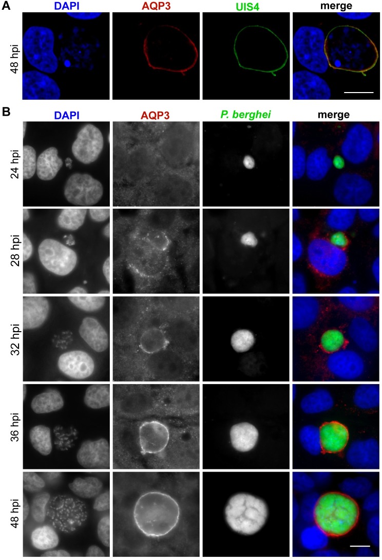
Fig 2. AQP3 localizes to the PVM in P. berghei infected hepatocytes.
(A) Representative confocal image of a HepG2 cell infected with P. berghei and stained for UIS4 (green), HsAQP3 (red), and DAPI (blue) at 48 hpi. Images represent 0.8 μm sections, scale bar = 10 μm. Pearson colocalization coefficient of 0.556 ± 0.014 (mean ± SD) calculated from the z-stacked images of three individual cells stained for HsAQP3 and UIS4. (B) Widefield fluorescent microscopy of HuH7 cells infected with GFP-expressing P. berghei (green) and fixed at various times post infection. Cells were stained for HsAQP3 (red) and DAPI (blue). Earliest localization of AQP3 to the PVM was detected at 28 hpi. After 32 hpi all EEFs have AQP3 staining. Scale bar, 10 μm.

