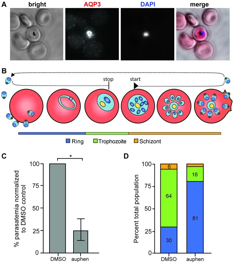
Fig 6. AQP3 localizes to the PVM and auphen treatment delays P. falciparum development in erythrocytes.
(A) Fluorescent microscopy of erythrocytes infected with P. falciparum 3D7. Cells were stained with HsAQP3 (red) and Hoechst (blue). AQP3 localizes to the PV of infected cells. (B) Schematic for auphen treatment of P. falciparum infected erythrocytes. Cells were treated with 2 μM auphen at the early schizont stage for 40 hours. (C) Parasitemia in auphen-treated cultures decreased 75 ± 12% compared to DMSO treated P. falciparum-infected erythrocytes (*p = 0.0257, unpaired Student’s t-test; n = 3 independent experiments). (D) After 40 hours of auphen treatment parasites were scored as ring, trophozoite, or schizont parasites. Auphen-treated P. falciparum exhibited a greater population of ring stage parasites when compared to the DMSO-treated control (n = 1 independent experiment).

