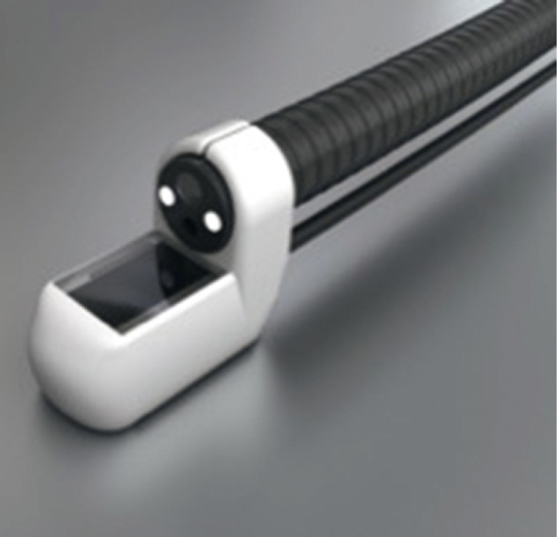
Fig. 1.

EndoVE (source: Cork Cancer Research Centre). Images of the device used in the trial- EndoVE ® . The electrode is attached to an endoscope. Tumor tissue is captured in place within the chamber (1 × 1 × 1.5 cm) of the device by a vacuum; this brings the tumor in contact with the two parallel electrodes which deliver pulses in 100 µs intervals. The procedure is repeated until the whole tumor area is covered.
