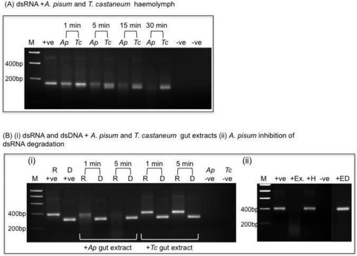
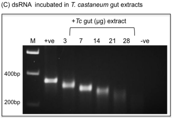
Figure 6.
In vitro stability of dsRNAs. (A) dsRNA (200 ng) incubated in the presence of A. pisum (Ap) or T. casteneum (Tc) cell free haemolymph (25 µg protein); −ve controls are Ap or Tc haemolymph alone. (B) (i) 200 ng dsRNA (R) or dsDNA (D) incubated with 3 µg Ap or Tc gut extract for 1 and 5 min. +ve denotes dsRNA and dsDNA control (i.e., no added protein), −ve control is gut extract alone. (B) (ii) Inhibition of dsRNA degradation in Ap gut extract. Samples were incubated for 5 min at 25 °C; +ve control is dsRNA in MOPS buffer; +Ex is dsRNA incubated with 3 µg Ap gut protein; +H is dsRNA incubated with heat treated (65 °C for 10 min) Ap gut extract (3 µg protein); +ED is dsRNA incubated with gut extract in MOPS buffer with 20 mM EDTA. (C) dsRNA (200 ng) incubated for 30 min with increasing amounts of T. castaneum gut protein (as denoted).


