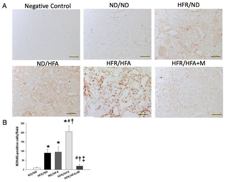
Figure 4.
(A) Light microscopic findings of 8-hydroxydeoxyguanosine (8-OHdG) immunostaining in the kidney cortex in 12-week-old male offspring. Bar = 50 μm; (B) Quantitative analysis of 8-OHdG-positive cells per microscopic field (400×); * p < 0.05 vs. ND/ND, # p < 0.05 vs. HFR/ND, † p < 0.05 vs. ND/HFA, ‡ p < 0.05 vs. HFR/HFA.

