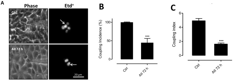
Figure 5.
AngII decreases gap junctional coupling between mesangial cells. (A) Etd+ was microinjected into the brightest cell (arrow) and diffused to neighboring cells. The right panels show fluorescence of Etd+ at different times (0 and 72 h) and the left panels are the phase contrast images. Coupling incidence (B) and coupling index (C) were evaluated in confluent mesangial cell cultures with AngII for different time periods (0 and 72 h) using dye (Etd+, 25 μM) coupling technique (black bars). Each bar represents the mean value ± SE of 4 independent experiments. In each experiment the dye was microinjected into at least 10 cells. Statistical significance *** p < 0.001 vs. Control. Scale bar = 20 μm.

