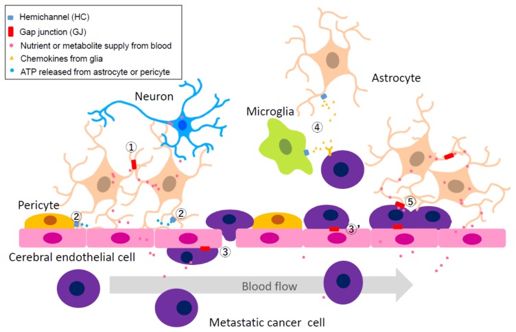
Figure 2.
The peri-vascular niche of the tumor microenvironment highlighting the extravasation of metastatic cells. The neurovascular unit includes cerebral endothelial cells (CECs), pericytes, glial cells, and neurons. Metastatic cancer cells utilize the nutrient supply from CECs to CNS cells mediated by the astrocytes. Numbers in circle indicate specific communication pathways. Pathway 1: GJ between normal astrocytes; it supports nutrients from CECs to CNS neurons. Pathway 2: HC activity of astrocytes and pericytes; it adjusts [Ca2+]i concentration in nearby CECs via release of ATP which results in change of blood flow rate. Pathway 3: GJ between CEC and metastatic cancer cell inside the capillary; it supports extravascular liberation of cancer cell from blood vessels. Pathway 3′: GJ between CEC and metastatic cancer cell located in a pericyte-like location; it protects cancer cells from immune attack by CNS astrocyte and microglia. Pathway 4: HC activity of microglia or astrocyte; it supports cancer growth by release of chemokines, although the actual mechanisms by which glial cells promote immune attack or support cancer remains unknown. Pathway 5: GJ between metastatic cancer cell located in a pericyte like-location and an astrocyte; it contributes to cancer growth as the first step in brain metastasis.

