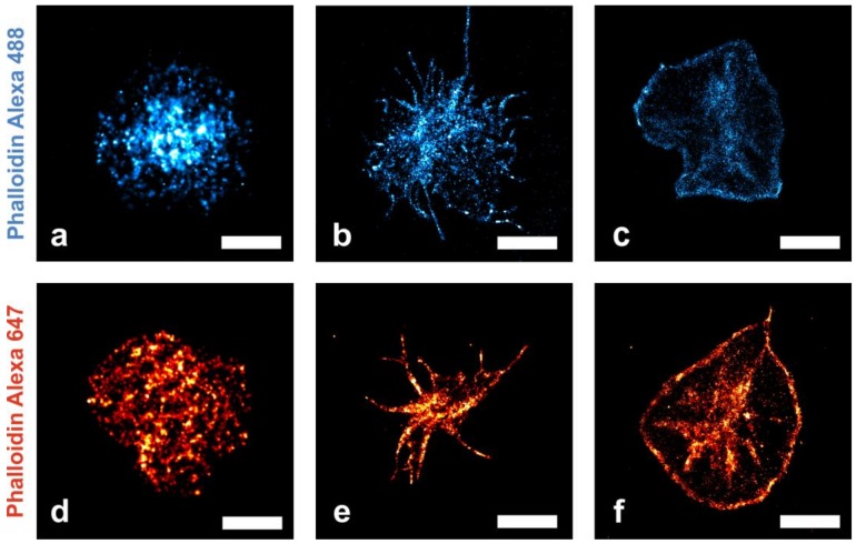
Figure 2.
Reconstructed fluorescence microscopy images (dSTORM) of human platelets at three different morphological states (cell culture medium on plain glass support), stained with phalloidin Alexa Fluor 488 (top row) or phalloidin Alexa Fluor 647 (bottom row). F-actin molecules bound by fluorescently labeled phalloidin are represented by the red and blue colored localizations within the cells, respectively. Brighter areas are caused by a higher density of localized fluorophores. As platelets start to adhere to the glass support, an early, round-shaped appearance (a,d) transforms into a spindle-like morphology (b,e) and subsequently yields a “fried-egg” shape (c,f). The central granulomere and accumulated actin in the peripheral cell region in the ‘fried-egg’ morphology is clearly visible (Figure 2c,f) [1]. Scale bar for (a,d) 1 µm and for (b,c,e,f) 3 µm.

