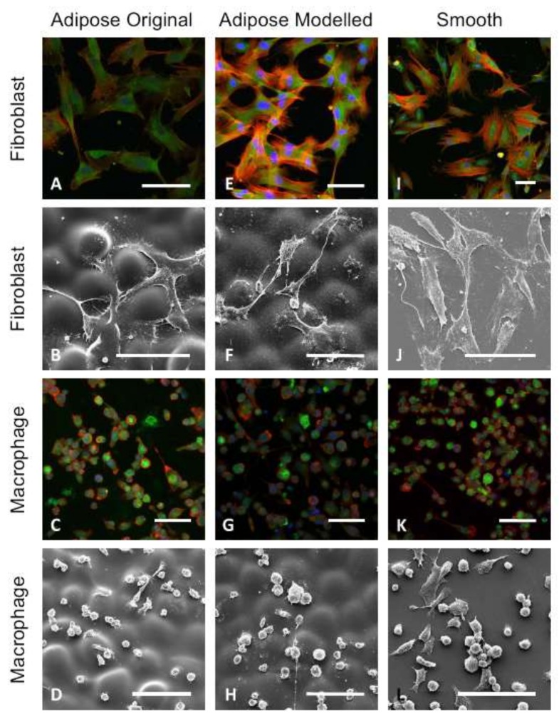
Figure 10.
Immunocytochemistry and SEM images (scale bars = 100 µm). Fibroblasts and macrophages were grown in culture on (A–D) the original adipose; (E–H) modeled adipose; and (I–L) smooth control surfaces; (A,E,I) Few differences were observed in focal contacts and cells were well spread and had a classical spindle shape; (B) Fibroblasts on the original adipose surface aligned with valleys between the hemispherical shapes; (F) while cells on the modeled surface spread across the surface and their secondary texture masked the primary hemispherical nature; (D,H) Macrophages did not align with the underlying primary topography and adhered to the uppermost surfaces of the original and modeled adipose surfaces; (D,H,L) Macrophages cultured on the modeled adipose surfaces spread less than those on the original adipose and smooth surfaces [120].

