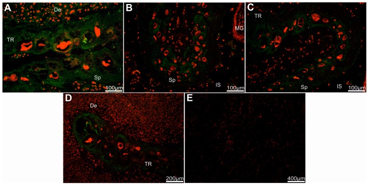
Figure 4.
Detection of the beaver pep/PAG-L within subplacenta sections. Immuno-localization of pep/PAG-L signals within subplacenta sections, identified by htdF-IHC with anti-pPAG-P (A) or anti-Rec pPAG2 polyclonals (B–D), visualized by goat anti-rabbit IgG-conjugated with Alexa 488 dye (A488; green) among all placental cells with nuclei stained by propidium iodine (red). (E) Negative control with omitted primary antibodies and nuclei stained by propidium iodine. Abbreviations: De—decidua; TR—trophectoderm; Sp—spongiotrophoblast; IS—intervillous space; MG—maternal gland.

