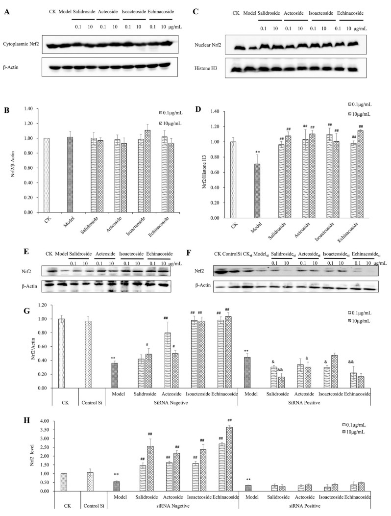
Figure 5.
Protective effect of PhGs on Nrf2 in H2O2-treated PC12 cells. (A) The expression of Nrf2 protein in cytoplasm was detected by immunoblotting using specific antibody. β-Actin was used as loading control. (B) The quantitative densitometric analysis of Nrf2 protein in cytoplasm. (C) The expression of Nrf2 protein in the nucleus was detected by immunoblotting using specific antibody. Histones H3 was used as loading control. (D) The quantitative densitometric analysis of Nrf2 protein in the nucleus. (E,F) PC12 cells were preincubated without (E) or with (F) Nrf2 siRNA for 24 h, then incubated with or without PhGs (0.1, and 10 μg/mL) for 24 h, and incubated with H2O2 for another 2 h after the PhGs were removed. Total Nrf2 protein expression was detected by immunoblotting using specific antibody, and β-actin was used as loading control. (G) The quantitative densitometric analysis of total Nrf2 protein. (H) The quantitative analysis of Nrf2 mRNA. (I) PC12 cells were preincubated with or without siRNA for 24 h, then incubated with or without PhGs (0.1 and 10 μg/mL) for 24 h, and incubated with H2O2 for another 2 h after the PhGs were removed. After treatment, the survival cells were determined by MTT assay. CK: normal group, Model: H2O2 treated group, ControlSi: control siRNA treated group, SiRNA Negative: without SiRNA treated group, SiRNA Positive: SiRNA treated group, Salidroside: salidroside treated group, Acteoside: acteoside treated group, Isoacteoside: isoacteoside treated group, Echinacoside: echinacoside treated group. ** p < 0.01 versus untreated group; # p < 0.05, versus H2O2 treated group (without Nrf2 siRNA treated), ## p < 0.01, versus H2O2 treated group (without Nrf2 siRNA treated); & p < 0.05 versus H2O2 treated group (with Nrf2 siRNA treated), && p < 0.01, versus H2O2 treated group (with Nrf2 siRNA treated).


