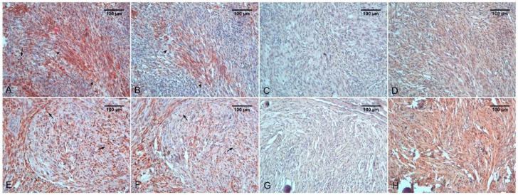
Figure 2.
Atypical meningioma (A–D). Immunostaining for CD68 (A), CD163 (B), iNOS (C), and HIF-1α (D). Note a CD68 positive signal in sheet-like growth areas (arrowheads) and in scattered macrophages (arrow). CD163 positivity is lower and immunoreactivity for HIF-1α is very low. Meningothelial meningioma (E–H). Immunostaining for CD68 (E), CD163 (F), iNOS (G), and HIF-1α (H). A more marked positivity for CD163 is evident. Note that CD68 positivity into the whorl is more marked than CD163 staining (Arrows) and strong positivity for HIF-1α.

