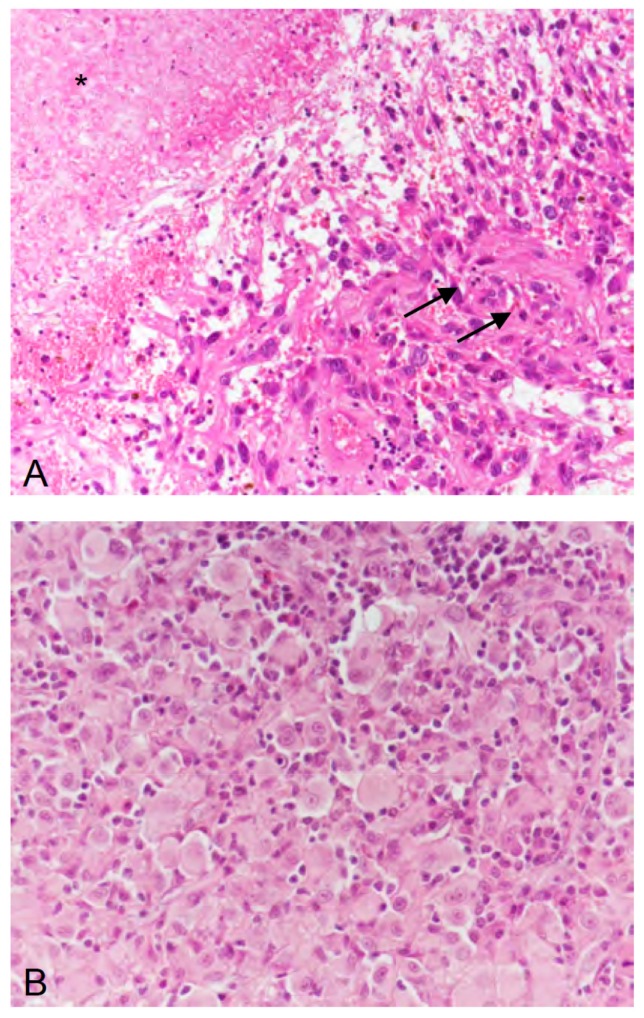
Figure 3.
(A) In 2007, the second recurrence of the tumor exhibited microvascular proliferation (arrows) and necrosis (asterisk), thus, corresponding to a glioblastoma (WHO grade IV). (B) In 2014, the fifth recurrence of the tumor was dominated by epithelioid differentiated glial tumor cells, thus, corresponding, to epithelioid glioblastoma (WHO grade IV). (A,B) hematoxylin and eosin staining; original magnification ×400.

